Riddles in code; is there a gene for language?

‘I have…’

Words are like genes; on their own they are not very powerful.  But apply them with others in the right phrase, at the right time and with the right emphasis, and they can change everything.

‘I have a dream…’

Genes are coded information.  They are like the words of a language, and can be combined into a story which tells us who we are.

The stories we choose to tell are powerful; they can change who we become, and also change the people with whom we share them.

‘I have a dream today!’


Language is a means for coding and passing on information, but it is cultural, and definitely non-genetic.  Nevertheless, for our speech capacity to have evolved, our ancestors must have had a body equipped to make speech sounds, along with the mental capacity to generate and process this language ‘behaviour’.  Our body’s development is orchestrated through the actions of relevant genes.  If the physical aspects of language ultimately have a genetic basis, this implies that speech must derive, at least in part, from the actions of our genes.

The hunt for genes involved with language led researchers at the University of Oxford to investigate an extended family (known as family KE).  Some family members had problems with their speech.  The pattern of their symptoms suggested that they inherited these difficulties as a ‘dominant’ character, and through a single gene locus.

The FoxP2 gene encodes for the ‘Forkhead-Box Protein-2’; a transcription factor.  This is a type of protein that interacts with DNA (shown here as a pair of brown spiral ladders), and influences which genes are turned on in the cell, and which remain silent.   This diagram shows two Forkhead box proteins, which associate with each other when active.  This bends the DNA strand and makes critical areas of the genetic code more accessible (Image: Wikimedia Commons)

The FOXP2 gene encodes for the ‘Forkhead-Box Protein-2’; a transcription factor. This is a type of protein that interacts with DNA (shown here as a pair of brown spiral ladders), and influences which genes are turne... mored on in the cell, and which remain silent. This diagram shows two Forkhead box proteins, which associate with each other when active. This bends the DNA strand and makes critical areas of the genetic code more accessible (Image: Wikimedia Commons)

Discovery of another unrelated patient with the same symptoms confirmed that the condition was linked to a gene known as FOXP2  (short for ‘Forkhead Box Protein-2’).  This locus encodes a ‘transcription factor’; a protein that influences the activation of many other genes.  FOXP2 was subsequently dubbed ‘the gene for language’.  Is that correct?

Not really.  FOXP2 affects a range of processes, not just speech.  The mutation which inactivates the gene causes difficulties in controlling muscles of the face and tongue, problems with compiling words into sentences, and a reduced understanding of language.  Neuroimaging studies showed that these patients have reduced nerve activity in the basal ganglia  region of the brain.  Their symptoms are similar to some of the problems seen in patients with debilitating diseases such as Parkinson’s and Broca’s Aphasia; these conditions also show impairment of the basal ganglia.

Genes code for proteins by using a 3-letter alphabet of adenine, thymine, guanine and cytosine (abbreviated to A, T, G and C).  These nucletodes are knwn as ‘bases’ (are alkaline in solution) and make matched pairs which form the ‘rungs of the ladder’ of the DNA helix. Substituting one base for another (as happens in many mutations) can change the amino acid sequence of the protein a gene encodes.  Changes may make no impact on survival, allowing the DNA sequence to alter over time.  Changes that affect critical sections of the protein (e.g. an enzyme’s active site), or critical proteins like FoxP2, are rare (Image: Wikimedia Commons)

Genes code for proteins by using a 3-letter alphabet of adenine, thymine, guanine and cytosine (abbreviated to A, T, G and C). These nucletodes are knwn as ‘bases’ (are alkaline in solution) and make matched pairs w... morehich form the ‘rungs of the ladder’ of the DNA helix. Substituting one base for another (as happens in many mutations) can change the amino acid sequence of the protein a gene encodes. Changes may make no impact on survival, allowing the DNA sequence to alter over time. Changes that affect critical sections of the protein (e.g. an enzyme’s active site), or critical proteins like FOXP2, are rare (Image: Wikimedia Commons)

Genes provide the code to build proteins.  Proteins are assembled from this coding template (the famous triplets) as a sequence of amino acids, strung together initially like the carriages of a train and then folded into their finished form.  The amino acid sequences of the FOXP2 protein show very few differences across all vertebrate groups.  This strong conservation of sequence suggests that this protein fulfils critical roles for these organisms.  In mice, chimpanzees and birds, FOXP2 has been shown to be required for the healthy development of the brain and lungs.  Reduced levels of the protein affect motor skills learning in mice and vocal imitation in song birds.

The human and chimpanzee forms of FOXP2 protein differ by only two amino acids. We also share one of these changes with bats.  Not only that, but there is only one amino acid difference between FOXP2 from chimpanzees and mice.  These differences might look trivial but they are probably significant.  FOXP2 has evolved faster in bats than any other mammal, hinting at a possible role for this protein in echolocation.

Mouse brain slice, showing neurons from the somatosensory cortex (20X magnification) producing green fluorescent protein (GFP).  Projections (dendrites) extend upwards towards the pial surface from the teardrop-shaped cell bodies. Humanised Foxp2 in mice causes longer dendrites to form on specific brain nerve cells, lengthens the recovery time needed by some neurons after firing, and increases the readiness of these neurons to make new connections with other nerves (synaptic plasticity).  The degree of synaptic plasticity indicates how efficiently neurons code and process information (Image: Wikimedia Commons)

Mouse brain slice, showing neurons from the somatosensory cortex (20X magnification) producing green fluorescent protein (GFP). Projections (dendrites) extend upwards towards the pial surface from the teardrop-shaped ce... morell bodies. Humanised Foxp2 in mice causes longer dendrites to form on specific brain nerve cells, lengthens the recovery time needed by some neurons after firing, and increases the readiness of these neurons to make new connections with other nerves (synaptic plasticity). The degree of synaptic plasticity indicates how efficiently neurons code and process information (Image: Wikimedia Commons)

Changing the form of mouse FOXP2 to include these two human-associated amino acids alters the pitch of these animals’ ultrasonic calls, and affects their degree of inquisitive behaviour.  Differences also appear in their neural anatomy.  Altering the number of working copies (the genetic ‘dose’) of FOXP2 in mice and birds affects the development of their basal ganglia.

Mice with ‘humanised’ FOXP2 protein show changes in their cortico-basal ganglia circuits along with altered exploratory behaviour and reduced levels of dopamine (a neurotransmitter  that affects our emotional responses).  So too, human patients with damage to the basal ganglia show reduced levels of initiative and motivation for tasks.

This suggests that FOXP2 is part of a general mechanism that affects our thinking, particularly around our initiative and mental flexibility.  These are critical components of human creativity, and are as it happens, essential for our speech.

Basal ganglia circuits process and organise signals from other parts of the brain into sequences.  Speaking involves coordinating a complex sequence of muscle actions in the mouth and throat, and synchronising these with the out-breath.  We use these same muscles and anatomical structures to breathe, chew and swallow;  our ability to coordinate them affects our speech, although this is not their primary role.

Family KE’s condition, caused by a dominant mutation in the FoxP2 gene, follows an autosomal (not sex-linked) pattern of inheritance, as shown here.   Dominant mutations are visible when only one gene copy is present.  In contrast a recessive trait is not seen in the organism unless both chromosomes of the pair carry the mutant form of the gene.   The FoxP2 transcription factor protein is required in precise amounts for normal function of the brain.  The loss of one working FoxP2 gene copy reduces this ‘dose’ which is enough to cause the problems that emerged as family KE’s symptoms (Image: Annotated from Wikimedia Commons)

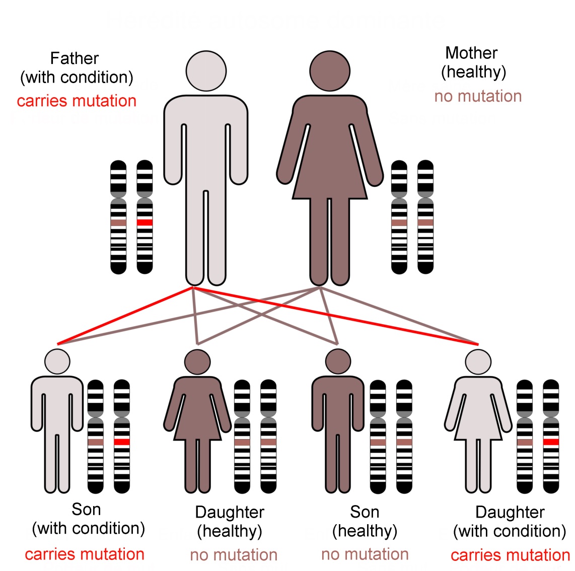
Family KE’s condition, caused by a dominant mutation in the FOXP2 gene, follows an autosomal (not sex-linked) pattern of inheritance, as shown here.Dominant mutations are visible when only one gene copy is present. In... more contrast a recessive trait is not seen in the organism unless both chromosomes of the pair carry the mutant form of the gene. The FOXP2 transcription factor protein is required in precise amounts for normal function of the brain. The loss of one working FOXP2 gene copy reduces this ‘dose’ which is enough to cause the problems that emerged as family KE’s symptoms (Image: Annotated from Wikimedia Commons)

In practice, very few of our 25,000 genes are individually responsible for noticeable characteristics.  Most genetically inherited diseases result from the effects of multiple gene loci.  FOXP2 is unusual because of its ‘dominant’ genetic character.  It does not give us our language abilities, but it is involved in the neural basis of our mental flexibility and agility at controlling the muscles of our mouths, throats and fingers.

In addition, genes are only part of the story of our development.  The way we think and subsequently behave alters our emotional state.  Feeling stressed or calm affects which circuits are active in our brain.  This alters the biochemical state of body organs and tissues, particularly of the immune system, modifying which genes they are using.

The dance between the code stored in our genes and the consequences of our thoughts builds us into what we are mentally, physically and socially.  This story is ours to tell.  By our experience, and with this genetic vocabulary, we create what we become.

Text copyright © 2015 Mags Leighton. All rights reserved.

References
Chial H (2008)  ‘Rare genetic disorders: Learning about genetic disease through gene mapping, SNPs, and microarray data’ Nature Education 1(1):192  http://www.nature.com/scitable/topicpage/rare-genetic-disorders-learning-about-genetic-disease-979
Clovis YM et al. (2012) ‘Convergent repression of Foxp2 3′UTR by miR-9 and miR-132 in embryonic mouse neocortex: implications for radial migration of neurons’  Development 139, 3332-3342.
Enard, W (2011) ‘FOXP2 and the role of cortico-basal ganglia circuits in speech and language evolution’  Current Opinion in Neurobiology  21; 415–424
Enard, W et al (2009)  A Humanized Version of Foxp2 Affects Cortico-Basal Ganglia Circuits in Mice  Cell 137 (5); 961–971  http://www.sciencedirect.com/science/article/pii/S009286740900378X
Feuk L et at., Absence of a Paternally Inherited FOXP2 Gene in Developmental Verbal Dyspraxia, in The American Journal of Human Genetics, Vol. 79 November 2006, p.965-72.
Fisher SE and Scharff C (2009) ‘FOXP2 as a molecular window into speech and language’  Trends in Genetics 25 (4); 166-177
Lieberman P  (2009)  ‘FOXP2 and Human Cognition’  Cell 137; 800-803
Marcus GF & Fisher SE (2003) ‘FOXp2 in focus; what can genes tell us about speech and language?’  Trends in Cognitive Sciences 7(6); 257-262
Reimers-Kipping S et al. (2011) ‘Humanised Foxp2 specifically affects cortico-basal ganglia circuits’ Neuroscience 175; 75-84
Scharff C & Haesler S (2005) ‘An evolutionary perspective on Foxp2; strictly for the birds?’ Current opinion in Neurobiology 15:694-703
Vargha-Khadem F et al. (2005) ‘FOXP2 and the neuroanatomy of speech and language’  Nature Reviews Neuroscience 6, 131-138 http://www.nature.com/nrn/journal/v6/n2/full/nrn1605.html
Wapshott N (2013)  ‘Martin Luther King's 'I Have A Dream' Speech Changed The World’ Huffington post, 28th August 2013  http://www.huffingtonpost.com/2013/08/28/i-have-a-dream-speech-world_n_3830409.html
Webb DM & Zhang J (2005) ‘Foxp2 in song learning birds and vocal learning mammals’  Journal of Heredity 96(3);212-216

Blind evolution gives eyeless fish sleepless nights

As the bats chatter above you in the cavern roof, their droppings rain down into the pool below.  The floor is silky with bacteria.  It’s cold and still.  A musty tang lingers in the air.  

You are completely blind.  You rely on hairs in your skin to feel for movement in the water. 

You are hungry.  Something is disturbing the mud; you can smell it!   You follow the scent trail and grab at it.  It feels like a shrimp. 

Things touch and nip you…  You are afraid of being eaten.  You move slowly, dozing for short periods, but don’t sleep. 

You have been here for…  How long?  Can you tell this without a sense of day and night?


The enhanced touch and taste senses of cave fish are also found in fish that hunt in murky waters, e.g. this channel catfish (Ictalurus punctatus).  These fish have touch sensitive ‘whiskers’ (barbels) and a high density of chemoreceptors (taste organs) over their bodies, making them into a ‘tactile, swimming tongue’.  These senses are more important to this fish than vision, hence their eyes are small (Image: Wikimedia Commons)

The enhanced touch and taste senses of cave fish are also found in fish that hunt in murky waters, e.g. this channel catfish (Ictalurus punctatus). These fish have touch sensitive ‘whiskers’ (barbels) and a high den... moresity of chemoreceptors (taste organs) over their bodies, making them into a ‘tactile, swimming tongue’. These senses are more important to this fish than vision, hence their eyes are small (Image: Wikimedia Commons)

Mexico’s blind cave fish show many of the adaptations found in animals from cave ecosystems across the world.  They have lost their eyes, and instead have chemoreceptors (‘taste buds’) scattered over their skin, allowing them to follow ‘pathways’ of chemical concentration (chemotaxis).  Touch-sensitive hairs around the mouth, along with a well-developed lateral line system, enable them to sense tiny water currents caused by prey and other fish.

In these caves, as in the deep sea, food is always in short supply.  To avoid being eaten by other fish they must remain vigilant, and so may ‘doze’ but do not sleep.  These fish have a low metabolic rate to conserve energy, and build up reserves of body fat .

Why do these fish and other cave dwellers go blind? One explanation is that eye loss is neutral to their survival.  When a characteristic is no longer essential to survival, mutations (mistakes in the DNA) that cause crucial genes to cease working are not selected against.  They are then passed to the next generation ‘at random’.

Random bead sampling experiments show how certain forms of a gene can become quickly widespread in a few generations in a small isolated population. This is known as ‘genetic drift’ (Image: Wikimedia Commons)

Random bead sampling experiments show how certain forms of a gene can become quickly widespread in a few generations in a small isolated population. This is known as ‘genetic drift’ (Image: Wikimedia Commons)

These small populations of cave fish were likely founded from only a few individuals.  We could anticipate that neutral mutations present in these founders would become widespread in the population by chance.  However these cave fish have evolved separately multiple times.  If the loss of eyes and skin pigment were a random, neutral process, we could expect some of these populations to have retained their sight and colour.  Also the PAX6 gene, used to build eyes in nearly all animals, is present and working in these fish.

Another possibility is energy conservation.  Eyes are costly to build and maintain, so disposing of them in this energy-poor environment seems a sensible option.  This however doesn’t fit the facts.  Eye cups are present in day-old embryos.  By day two, the lens cells are beginning to grow and divide, but in these cave fish a process of deliberate ‘cell death’ destroys them as they arise.  This strategy seems neither neutral nor particularly economic.

So what is really driving the evolution of these pale, sightless and sleepless fish?  Genetic studies are providing some alternative answers that shine light into how evolution really works.

Why do cave fish kill off their own eyes? 

A cross-section of a mouse eye showing the PAX6 protein, visible here in green thanks to a fluorescent  tag.   PAX6 is a transcription factor; a protein that controls the actions of many other genes.  Its biological role is to define which cells develop into eyes in all vertebrate embryos.   The PAX6 gene functions in the Mexican blind cave fish, but works at less intensity than in sighted fish.   Cave fish eyes develop normally for the first 2 days in the embryo, then stop growing, and the cells degenerate.  These fish appear eyeless because other tissues expand and cover what remains of the eye cups (Image: Wikimedia Commons)

A cross-section of a mouse eye showing the PAX6 protein, visible here in green thanks to a fluorescent tag. PAX6 is a transcription factor; a protein that controls the actions of many other genes. Its biological role is... more to define which cells develop into eyes in all vertebrate embryos. The PAX6 gene functions in the Mexican blind cave fish, but works at less intensity than in sighted fish. Cave fish eyes develop normally for the first 2 days in the embryo, then stop growing, and the cells degenerate. These fish appear eyeless because other tissues expand and cover what remains of the eye cups (Image: Wikimedia Commons)

The PAX gene plays a key role in the development of eyes in vertebrate embryos.  PAX6 action is reduced by increases in the activity of another gene, called HEDGEHOG whichdefines boundaries between cell types and promotes development of the fish’s lateral line, jaws, teeth and taste buds.  HEDGEHOG is more active in cave fish embryos than in their sighted sister species, causing the lateral line cell zone and the jaw to expand.  As it does so, this also switches off PAX6 and halts eye development.

This trade-off between ‘touch and taste’ versus ‘sight’ enhances the very senses that the cave fish needs in its ever-dark world.  Eye shrinkage, which reduces the energy budget, is a secondary effect of selection for these other senses.  This subtle change in the key interaction of two developmental genes  at a critical stage can radically alter a gene’s effect and the body which results.

What does enhanced touch sensitivity provide for these fish?

Try stroking your eyebrow against the direction of hair growth. Now stroke your forehead. Which feels like a stronger movement?

A school of sardines shoaling together with precise coordination.   Fish have an awareness of space that is provided by their lateral line system.  This sensory mode is able to pick up changes in turbulence around rocks and other obstacles, and to detect the movements of other fish.  This is how shoals are able to swim together without colliding (Image: Wikimedia Commons)

A school of sardines shoaling together with precise coordination.Fish have an awareness of space that is provided by their lateral line system. This sensory mode is able to pick up changes in turbulence around rocks and... more other obstacles, and to detect the movements of other fish. This is how shoals are able to swim together without colliding (Image: Wikimedia Commons)

Hairs in our skin enable us to be more sensitive to touch.  The Mexican blind cave fish have a lateral line systemlike all fish.  All lateral line systems use tiny hair cells to detect changes in water movement, but in these cave dwellers it is unusually sensitive.

These fish also have a behaviour which seems counter-intuitive.  If introduced into a new environment, instead of slowing down and moving cautiously, they swim faster.  The reason is that more rapid movements increase the flow of information to the hair cells.  This expands their awareness, providing a larger ‘hydrodynamic image’ of their world.  Also at greater speeds, the water layer that clings to their skin is reduced, helping to make this image ‘sharper’.

Why are cave fish so pale?

Cave animals typically lose their colour and cease to develop eyes, like this Texas Blind Salamander (Eurycea rathbuni).   Cave ecosystems have limited air exchange with above ground.  Often the air is low in oxygen.   This film clip shows the salamander’s external gills, a juvenile characteristic retained here in the adult form in order to capture enough oxygen gas (Image: Wikimedia Commons)

Cave animals typically lose their colour and cease to develop eyes, like this Texas Blind Salamander (Eurycea rathbuni). Cave ecosystems have limited air exchange with above ground. Often the air is low in oxygen. This ... morefilm clip shows the salamander’s external gills, a juvenile characteristic retained here in the adult form in order to capture enough oxygen gas (Image: Wikimedia Commons)

The melanin-based pigments in our skin and those of other animals provide colour and pattern, but they have a more ancient evolutionary role: to  protect us from damage by ultraviolet light.  Without sunlight, cave animals typically are colourless.  In Mexican cave fish this is connected to a loss-of-function mutation (in the gene oca2), controlling the first step of pigment production.

Independently evolved populations of cave fish are colourless thanks to unique mutations in this same gene.  This is curious.  If pigment loss occurred by chance, we would expect that some populations would have shut down later stages of pigment production, as this would give the same effect.  This specific mutation suggests instead that pigment loss has been actively selected.

Pigment loss may conserve energy, and it is possible that mutating the genes controlling later biosynthetic stages may have other effects that reduce fitness.  However a more compelling explanation is that shutting down oca2 increases the availability of tyrosine, an amino acid.  What is so special about tyrosine?

Panellus stipticus is a fungus that grows on dead wood. The bioluminescent strain is nicknamed "glow wood" (Image: Wikimedia Commons)

Panellus stipticus is a fungus that grows on dead wood. The bioluminescent strain is nicknamed “glow wood” (Image: Wikimedia Commons)

Neurotransmitter production depends upon the tyrosine supply.  The fish produces the neurotransmitters dopamine and noradrenaline (norepinephrine), and the hormone adrenaline (epinephrine), from tyrosine.  Cavefish brains have higher concentrations of these chemicals than brains of sighted fish.  Noradrenaline and adrenaline provoke the ‘fight or flight response; in these fish they are linked to the minimal amounts of sleep and the ability to engage in unusually fast foraging.

Adaptation to life in caves has produced a range of animals with some remarkably similar characteristics.  It seems that Mexico’s cave fish have made an evolutionary trade-off.  Faster swimming and an enhanced sensitivity to touch and taste comes at the cost of eyes and skin colour.  And it keeps them awake in the dark.

Text copyright © 2015 Mags Leighton. All rights reserved.

References
Bibliowicz, J, et al. (2013)  Differences in chemosensory response between eyed and eyeless Astyanax mexicanus of the Rio Subterráneo cave.  EvoDevo 4, e25.
Bilanžija, H. et al. (2013)  A potential benefit of albinism in Astyanax cave fish: down-regulation of the oca2 gene increases tyrosine and catecholamine levels as an alternative to melanin synthesis.  PLoS ONE 8, e80823.
Burt de Perera, T. and Braithwaite, V.A.  (2005)  Laterality in a non-visual sensory modality — the lateral line of fish.  Current Biology 15, R241-R242.
Coombs, S. et al. (2000)  Hydrodynamic image formation by the peripheral lateral line system of the Lake Michigan mottled sculpin, Cottus bairdi.  Philosophical Transactions of the Royal Society of London, B 355, 1111-1114.
Ćurčić-Blake, B. and van Netten, S.M. (2006)  Source location encoding in the fish lateral line canal.  Journal of Experimental Biology 209, 1548-1559.
Horstkotte, J. et al. (2010)  Predation by three species of spiders on a cavefish (Poecilia mexicana, Poeciliidae) in a Mexican sulphur cave.  Bulletin of the British Arachnological Society 15, 55-58.
Jeffery, W.R. (2001)  Cavefish as a model system in evolutionary developmental biology.  Developmental Biology 231, 1–12.
Jeffery, W.R. (2009)  Regressive evolution in Astyanax cave fish.  Annual Review of Genetics 43, 25-47.
Jeffery, W.R. et al. (2003)  To see or not to see: evolution of eye development in Mexican Blind Cavefish.  Integrative Comparative Biology 43, 531–541.
Mueller, K.P. et al. (2014)  Sunscreen for fish: the co-option of UV light protection for camouflage.  PLoS ONE 9, e87372.
Niven, J.E. (2008)  Evolution: convergent eye losses in fishy circumstances.  Current Biology 18, R27–R29.
Rétaux, S. and Casane, D. (2013)  Evolution of eye development in the darkness of caves: adaptation, drift, or both?  EvoDevo 4, 26.
Sanford, W.E. et al. (2006)  Research Opportunities in Interdisciplinary Ground-Water Science in the U.S. Geological Survey.  Circular 1293. U.S. Geological Survey.
Strickler, A.G. et al. (2007)  The lens controls cell survival in the retina: evidence from the blind cavefish Astyanax. Developmental Biology 311, 512-523.
Tian, N.M. and Price, D.J. (2005)  Why cavefish are blind. BioEssays 27, 235-238.
Wilkens, H. and Strecker, U. (2003)  Convergent evolution of the cavefish Astyanax (Characidae, Teleostiei): genetic evidence from reduced eye-size and pigmentation.  Biological Journal of the Linnean Society 80, 545–554.
Wilkens, H. (1988)  Evolution and genetics of epigean and cave Astyanax fasciatus (Characidae, Pisces).  Evolutionary Biology 23, 271–367.
Yamamoto, Y. et al. (2004)  Hedgehog signalling controls eye degeneration in blind cavefish.  Nature 431, 844-847.